

What Can — and Can't — a Panoramic Dental X-ray Show?
Short answer
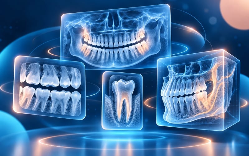
A panoramic X-ray (often called a “panoramic” or OPG) shows all your teeth and both jaws in a single wide image. It is good for an overview: missing, impacted or developing teeth, existing fillings and crowns, larger changes in the jawbone and the general bone level.
It is less detailed than small X-rays taken inside the mouth, so it can miss early decay between teeth or small changes at a root tip, and blur or positioning can create misleading shadows. Your dentist combines it with an examination and, when needed, other images.
On this page
What a panoramic X-ray is
For a panoramic X-ray you stand or sit in a machine that rotates around your head for a few seconds. The result is one flat, wide image of the upper and lower jaws, including all teeth, the surrounding bone and parts of nearby structures such as the jaw joints and the floor of the sinuses. It is taken from outside the mouth, so nothing has to be held between the teeth apart from a small bite block.
Dentists often use it at a first visit, before wisdom tooth removal, when planning larger treatment, or to follow children's developing teeth. Whether you need one depends on your history and examination; the American Dental Association recommends taking X-rays only when the dentist expects them to provide necessary diagnostic information.
What it shows well
- The big picture. How many teeth you have, which are missing, and how they are positioned.
- Wisdom teeth and impacted teeth. Teeth that have not come through, and their position relative to neighbouring teeth.
- Developing teeth in children and teenagers.
- Existing dental work — fillings, crowns, bridges, root canal fillings and implants usually show up clearly.
- Larger changes in the jaw, such as bigger areas around root tips or other changes in bone that need further investigation.
- The general bone level along the teeth, as an overview.
What it can miss
Because a panoramic image covers a large area, each tooth is shown with less fine detail than on X-rays taken inside the mouth. In practice that means:
- Early decay between teeth is often hard to see; guidance on dental radiography recommends bitewing X-rays for detecting decay.
- Small changes at a root tip or detailed root anatomy may need a periapical X-ray.
- Front teeth can look blurred, because this region is narrow in the image “slice” and sensitive to head position.
- Gum health cannot be judged from an X-ray: bleeding, pocket depth and gum recession are checked in the mouth.
- Symptoms — pain, sensitivity, a cracked tooth that hurts on biting — often have no clear sign on any X-ray.
A panoramic image is also two-dimensional, so it cannot show the exact width of bone in three dimensions. Our guide to panoramic, bitewing, periapical and CBCT images explains when each is used.
Why some panoramic images are misleading
Panoramic X-rays are sensitive to how the head is positioned. A review of common positioning errors describes, for example, a dark band over the upper teeth when the tongue is not resting against the palate, front teeth that look too wide or too narrow when the head is too far forward or back, and a “smile” or “frown” shape when the chin is tilted. Moving during the scan, earrings, glasses or a protective thyroid collar in the beam path can also leave shadows.
These artefacts matter for both people and software: an overlapping shadow can look like decay, and a blurred area can hide a real change. If an image has obvious problems, your dentist may judge that it cannot answer a particular question.
What happens next
Your dentist reads the panoramic image together with your history and an examination. If a question remains — for example, a suspicious area between two back teeth — they may suggest a more detailed image for that region rather than repeating the whole overview. If you already have recent X-rays from another clinic, you can ask for copies; the ADA notes that a new dentist may be able to use previous images instead of taking new ones.
If you plan to share a panoramic image online, a few quality checks before uploading help make sure it shows what it should.
What this means for you
- Keep a copy of your panoramic X-ray, with the date it was taken.
- Expect your dentist to examine you as well; the image is not the whole assessment.
- If a problem is suspected, ask whether a more detailed image of that area would help.
- Do not ask for an X-ray simply to have one; your dentist weighs benefit against radiation exposure.
Frequently Asked Questions
Is a panoramic X-ray the same as an OPG?
Yes. OPG stands for orthopantomogram, another name for a panoramic dental X-ray. You may also see “panoramic radiograph” or “panorex”.
Can a panoramic X-ray show cavities?
It can show larger areas of decay, but early decay between teeth is often missed. Bitewing X-rays are the usual choice when a dentist is looking for decay between back teeth.
Does a panoramic X-ray show gum disease?
It can show the overall bone level around teeth, which is one sign used in assessing gum disease. The gums themselves — bleeding, pocket depth, inflammation — have to be examined in the mouth.
Is a panoramic X-ray enough to plan an implant?
It can help with an overview, but it is a two-dimensional image and cannot show bone width or nearby structures in three dimensions. Your dentist decides whether additional imaging, such as CBCT, is needed.
How often should I have one?
There is no fixed schedule. It depends on your age, risk, symptoms and treatment needs, and your dentist decides when the benefit justifies a new image.
Sources and further reading
- X-Rays/Radiographs (Oral Health Topics) (opens in a new tab)
- X-rays (opens in a new tab)
- Selection Criteria for Dental Radiography (3rd edition) (opens in a new tab)
- Common positioning errors in panoramic radiography: A review (opens in a new tab)
Reviewed on