

Panoramic, Bitewing, Periapical and CBCT: What's the Difference?
Short answer
Each type of dental image answers a different question. A panoramic X-ray gives an overview of both jaws; a bitewing shows the crowns of the back teeth and is the usual choice for decay between them; a periapical shows one or a few teeth from crown to root tip; and CBCT is a three-dimensional scan used for specific problems such as implant planning or complex roots.
No single image shows everything, and more imaging means more radiation, so dentists choose the image that answers the clinical question with the lowest reasonable exposure.
On this page
Panoramic (OPG): the overview
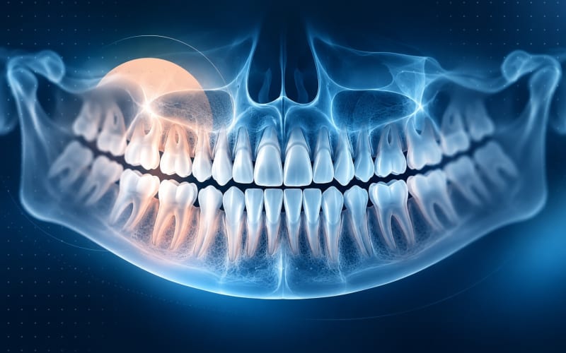
A panoramic X-ray is taken outside the mouth while the machine rotates around your head. It shows both jaws, all teeth and surrounding bone in one wide, flat image.
Typically used for: a first overview, wisdom teeth and impacted teeth, developing teeth in children, larger changes in the jaw, and as a starting point for planning.
Limits: less fine detail than images taken inside the mouth, possible blur in the front teeth, and sensitivity to head position. Read more in what a panoramic X-ray can and can't show.
Bitewing: decay between the back teeth
For a bitewing, you bite on a small tab or holder with the sensor inside your mouth. Each image shows the crowns of the upper and lower back teeth on one side, plus the bone level between them.
Typically used for: detecting decay between back teeth, which is often invisible to the eye; checking the edges of fillings; and following bone levels over time. Selection-criteria guidance treats bitewings as the main radiograph for caries detection.
Limits: roots and root tips are usually not fully visible, and front teeth are not included.
Periapical: one tooth, root to tip
A periapical X-ray is also taken with a sensor inside the mouth, but positioned to capture one or a few teeth in full — from the crown down to the root tip (the apex) and the bone around it.
Typically used for: a painful or sensitive tooth, suspected infection at the root tip, root canal treatment and its follow-up, injuries to teeth, and checking an implant or a tooth before treatment.
Limits: it covers a small area, and like all 2D images it can hide what lies in front of or behind the tooth.
CBCT: three dimensions, for specific questions
Cone beam computed tomography (CBCT) produces a three-dimensional image of a selected area of the jaws. It lets a dentist look at bone and teeth in slices from any direction.
Typically used for: implant planning (bone height and width, nearby nerves and sinuses), the position of impacted teeth close to nerves, complex root canal anatomy or problems that 2D images could not explain, and some bone and jaw-joint questions.
Limits: the radiation dose is higher than for standard 2D dental X-rays, metal fillings and implants can cause streak artefacts, and soft tissues such as gums are not shown well. Professional statements — including the ADA's advisory on CBCT and the European Society of Endodontology's position on CBCT in root canal treatment — advise using it when conventional imaging cannot answer the clinical question, not as a routine first image.
At a glance
| Image | Shows | Best for | Main limits |
|---|---|---|---|
| Panoramic | Both jaws, all teeth, in one 2D view | Overview, wisdom teeth, development, larger changes | Less detail; positioning artefacts |
| Bitewing | Crowns of upper and lower back teeth | Decay between teeth, filling edges, bone level | No root tips; back teeth only |
| Periapical | One or a few whole teeth, root tip included | Pain, root tip problems, root canal treatment | Small area; 2D overlap |
| CBCT | A 3D volume of a selected area | Implant planning, complex roots, impacted teeth near nerves | Higher dose; metal artefacts; used selectively |
Who decides which image you need
Choosing an image is a clinical decision. Your dentist considers your symptoms, history, age, risk of decay or gum disease, and any recent images, then picks the image that answers the question with the lowest reasonable radiation dose — the principle often summarised as ALARA (“as low as reasonably achievable”). The ADA encourages patients and dentists to discuss the need for X-rays together.
If you already have recent images, bring them: they may avoid a repeat. Online tools work with the images you have; they should never be a reason to take new ones. If you share images online, the upload quality checklist helps you send the most useful version.
What this means for you
- Ask your dentist which question an X-ray is meant to answer.
- Keep copies of your images with dates and the type of image.
- Mention previous X-rays or scans so they can be reused where appropriate.
- Don't expect one image to show everything; a combination of images and an examination may be needed.
Frequently Asked Questions
Which X-ray is best for finding cavities?
For decay between back teeth, bitewing X-rays are the usual choice. A dentist also checks teeth visually, and may use other images for specific teeth.
Is CBCT better than a normal dental X-ray?
It gives different information. CBCT shows three dimensions but uses a higher radiation dose and is reserved for questions that 2D images cannot answer, such as some implant planning or complex root problems.
Why does my dentist want a periapical X-ray when I already have a panoramic?
A periapical image shows one tooth in much more detail, including the root tip. It can answer a question about a painful tooth that the panoramic overview cannot.
Are dental X-rays safe?
Dental X-rays use low radiation doses, and dentists follow the principle of keeping exposure as low as reasonably achievable. Tell your dentist if you are or may be pregnant; they will take it into account.
Can I upload a CBCT scan for AI analysis?
Many online tools, including LUMBEN's patient upload, are designed for 2D dental radiographs. CBCT data are large 3D files that are usually reviewed by a dentist with specialised software.
Sources and further reading
- X-Rays/Radiographs (Oral Health Topics) (opens in a new tab)
- Selection Criteria for Dental Radiography (3rd edition) (opens in a new tab)
- The use of cone-beam computed tomography in dentistry: an advisory statement from the American Dental Association Council on Scientific Affairs (opens in a new tab)
- European Society of Endodontology position statement: Use of cone beam computed tomography in Endodontics (opens in a new tab)
- Dental Cone Beam CT (opens in a new tab)
Reviewed on